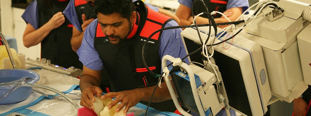
Dr. Iyer evaluates a 3D printed patient-specific heart model during a surgery dry run prior to treating the patient.
Physicians at the Jacobs Institute, a newly designated Stratasys-supported Center of Excellence, the University at Buffalo’s Clinical and Translational Research Center (CTRC) and Kaleida Health’s Vascular Institute in Buffalo, New York, have been relying on Stratasys’ PolyJet 3D printing solutions to develop treatment plans for life-threatening vascular issues such as aneurysms, stroke and blood clots. In addition to making exact models to match specific patients, Stratasys 3D Printers are being used to create pieces for medical training as well and to develop trial runs for new treatment protocols.
“We use 3D printing technology and materials to create a lifelike vascular environment that isn’t achievable any other way,” said Mike Springer, Director of Operations and Entrepreneurship at the Jacobs Institute.
Second-year medical students in training using 3D printed vascular models.
“3D printing is valuable in planning complex procedures with a team. Without it, we prepare for complications on a theoretical basis,” said Dr. Vijay Iyer, an interventional cardiologist. “Many times, despite the best theoretical planning, we are faced with circumstances where we don’t know what to do.”
Converting real patient-derived anatomy into realistic 3D models allows physicians to integrate visual and tactile clues into their surgical plan. Physical models allow the team to test theories and reveal potential complications before the patient is on the table and time is critical.
Free Webinar: Learn how 3D Printing can help you cost-effectively improve care, refine new medical devices and speed advances at every step in the medical value chain.
This 3D printed vascular testing model allows medical device designers and engineers to gather valuable performance feedback on device performance.
“3D vascular models represent a new paradigm for training the next generation of doctors. This paradigm includes surgical and endovascular simulation and skills evaluation before they are allowed to treat patients,” said Dr. L. Nelson Hopkins, founder of the Jacobs Institute and the Gates Vascular Institute.
3D printing enables the Jacobs Institute to accelerate and improve medical device design, and the team at CTRC also does preclinical testing for product validation using customized anatomical models to capture feedback on device performance.
“Recently, we tested how effectively a particular device could reach the brain depending on tortuosity of the anatomy. We designed a series of models with differing levels of tortuosity, then tested the devices,” said Dr. Adnan Siddiqui, chief medical officer at the Jacobs Institute, vice chairman and professor of neurosurgery at University at Buffalo Neurosurgery, and director of neurosurgical stroke service at Kaleida Health. “This is impossible to do in animals and patients, but 3D printing makes it easy in a smooth, streamlined process.”
In addition to improving new device performance, the early feedback that designers glean from real-anatomy testing lets them avoid costly, potentially unsuccessful animal testing.
Follow Stratasys Medical on our new LinkedIn Showcase page.
The Jacobs Institute, Kaleida Health’s Gates Vascular Institute and University at Buffalo’s Clinical and Translational Research Center use 3D printing as a platform for developing cutting-edge medical solutions.
Ciprian Ionita, Ph.D., is a research assistant professor of biomedical engineering and neurosurgery at University at Buffalo. His team at the Jacobs Institute regularly uses a 3D printer to create custom fixtures for scientific equipment and experiments. “Using our in-house Stratasys 3D printer, we don’t rely on external machine shops that would generate both lag time and expense. Most of these fixtures and components can be 3D printed within a few hours,” said Dr. Ionita.
Through its collaboration with GVI and CTRC, the Jacobs Institute is harnessing the power of a 3D printing platform in all aspects of health care.
Watch this video to learn how 3D printing is becoming an indispensable tool for the future of medicine!


